When Do Chromatids Become Chromosomes During Mitosis?
Many people are familiar with the word “chromosome” from high school biology class, but not everyone knows the details of how these structures form during the process of mitosis. Specifically, scientists have long been interested in understanding when chromatids become chromosomes during mitosis. This article aims to provide a comprehensive overview of this important event.
The purpose of this article is to educate readers on the details of the transition from chromatids to chromosomes in mitosis. It is intended for anyone interested in the basic mechanisms of cell division and the molecular biology of chromosomes and may be especially useful for high school or undergraduate students.
Mitosis Unraveled: Understanding Chromatids and Chromosomes
The process of mitosis is a key mechanism by which cells divide into two daughter cells. Mitosis occurs in somatic, or non-reproductive, cells and is responsible for the growth and repair of tissues in the body. Mitosis can be divided into four key stages: prophase, metaphase, anaphase, and telophase. During each of these stages, different processes occur within the cell that ultimately lead to division.
Chromosomes carry the genetic material of the cell and are essential for proper cell division. In humans, the typical somatic cell has 46 chromosomes, which are arranged in pairs. At the start of the mitotic process, each chromosome consists of a pair of identical structures, called chromatids, that are joined together at a central point called the centromere.
The distinction between chromatids and chromosomes is important. Chromatids are only visible during cell division, whereas chromosomes exist throughout the cell cycle. In other words, when the cell is not preparing to divide, chromatids are bound together and invisible to the naked eye. Only when the cell progresses through mitosis do these structures become more visible and recognizable as individual chromosomes.
A Step-by-Step Guide to Mitosis: When Do Chromatids Become Chromosomes?
Mitosis can be divided into four key stages, each of which has its own unique molecular events. During the early stages of mitosis, the cell’s chromatin (the loosely packed material that makes up the DNA and associated proteins) begins to condense into more compact structures. These more compact structures are the chromosomal pairs or, more specifically, the chromatid pairs.
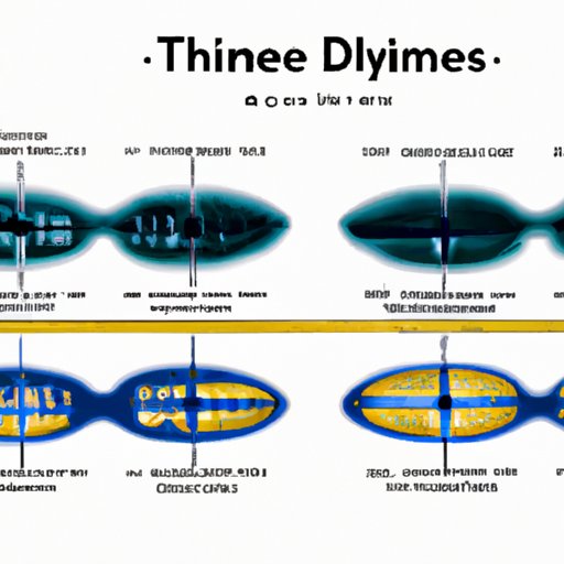
During the metaphase stage of mitosis, the pairs of chromatids align at the cell’s equator, known as the metaphase plate. This is the point at which the transition from chromatids to chromosomes begins. Once the chromatids are aligned at the metaphase plate, the centromeres of each chromatid pair split apart, freeing the individual chromatids to separate and migrate to opposite poles of the cell.
At this stage, each individual chromatid is now considered a separate chromosome because it carries its own complete set of genetic information. As the chromosomes are pulled apart and segregated towards opposite poles of the cell via the spindle fibers, they become more condensed and more visible under a microscope. By the end of mitosis, each of the eukaryotic daughter cells has an identical set of chromosomes in their nuclei.
The Crucial Transition: Chromatids to Chromosomes in Mitosis
The transition from chromatids to chromosomes is crucial because it sets the stage for the proper segregation of genetic material between daughter cells. This separation is important because each cell needs to have an identical set of chromosomes in order to function properly. Errors that occur during chromosomal segregation can lead to diseases such as Down syndrome, which is caused by an extra copy of chromosome 21.
Several factors contribute to the transition from chromatids to chromosomes. One important factor is the physical mechanics of the nucleus and spindle apparatus, which can apply stresses and forces to the chromatin and influence the progression of mitosis. Additionally, several proteins and protein complexes, such as the cohesin complex and the anaphase-promoting complex, are known to play important roles in the separation of chromatids during mitosis.
Mitotic Metamorphosis: The Transformation from Chromatids to Chromosomes
The transition from chromatids to chromosomes involves several changes in the structure and organization of the cell’s genetic material. When the chromatids first line up at the metaphase plate, they are still joined together via the centromere. As the centromeres divide, the individual chromatids become more exposed and visible as separate structures, each with their own complete set of genetic information.
As the individual chromosomes move towards opposite poles of the cell, they condense even further, forming compact, darkly-staining structures that are easily visible under a microscope. This increased level of compaction is thought to be driven by the activity of motor proteins, such as those found in the kinetochore region of the chromosome.
The Invisible Progression: Identifying Chromosome Formation during Mitosis
Scientists study the transition from chromatids to chromosomes in a variety of ways. One common method is to use fluorescently-labeled antibody probes that recognize specific proteins or DNA sequences within the chromatin. These probes can help researchers visualize the changes that occur during mitosis and track the progression of chromosomal segregation throughout the cell cycle.
More recent technological advances have allowed for the use of live-cell imaging techniques, which enable researchers to observe the behavior of mitotic cells in real-time. This has provided new insights into the molecular and physical processes that underlie the transition from chromatids to chromosomes during mitosis.
The Mystery Solved: Revealing the Mechanism of Chromosome Formation in Mitosis
Recent advances in molecular biology and live-cell imaging have shed new light on the transition from chromatids to chromosomes during mitosis. For example, researchers have identified key proteins that are involved in the separation of chromatids, such as the cohesin complex and the anaphase-promoting complex. Others have developed sophisticated imaging methods that have allowed them to observe the mechanical stresses and forces that are present during mitosis.
This improved understanding of the molecular and physical mechanisms of mitosis has important implications for biomedical research. Understanding how chromosomes are formed and segregated during cell division can help us gain new insights into the causes of genetic diseases and the development of new treatments for these conditions.
Conclusion
In conclusion, the transition from chromatids to chromosomes is a critical event during the process of mitosis. Understanding this process is essential for gaining insight into the fundamental mechanisms of cell division and the maintenance of genomic stability. By investigating the physical and molecular processes that underlie this transition, scientists are discovering new ways to treat genetic diseases and engineer new therapies in the future.
To learn more about this topic, readers may want to explore additional resources such as molecular biology textbooks and research papers.
